Knee
Anatomy
The knee is made up of two joints called the tibiofemoral joint and the patellofemoral joint. The tibiofemoral joint is a bony articulation between the tibia (lower leg) and femur (long thigh bone). The patellofemoral joint is a bony articulation between the patella (knee cap) and femur. An overview of the anatomy is shown in Figure 1.
- Ligaments are soft tissues that connect bone to bone and help to make the knee stable. There are four major ligaments associated with the knee joint: anterior cruciate ligament (ACL), posterior cruciate ligament (PCL), medial collateral ligament (MCL) and lateral collateral ligament (LCL). These ligaments work together with other soft tissues in the knee to control the amount of movement or laxity in your knee.
- Cartilage is rubber-like tissue that covers and protects the ends of long bones at the joints. There are two types of cartilage in your knee. Articular (or hyaline) cartilage lines your joint. The two menisci (medial and lateral) are fibrocartilage pads located in the joint space between the femur and tibia.
- Muscles that span the knee joint include the quadriceps, which helps to straighten your knee, and hamstrings, which help you to bend your knee. These muscles help you to move and make the knee stable. There are many other muscle groups in the lower limb and spine that will be involved in a rehabilitation programme and to help you move.
- Tendons are soft tissues that join the muscle to the bone. The patella and quadriceps tendons cross the front of the knee. The patella sits in the quadriceps tendon.
The following pictures showing the anatomy (bone and soft tissue structures) of the knee are taken by magnetic resonance imaging (MRI) of a knee with no injuries or knee symptoms.
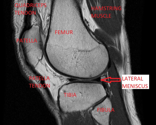
Figure 1: MRI image of the knee from the side.
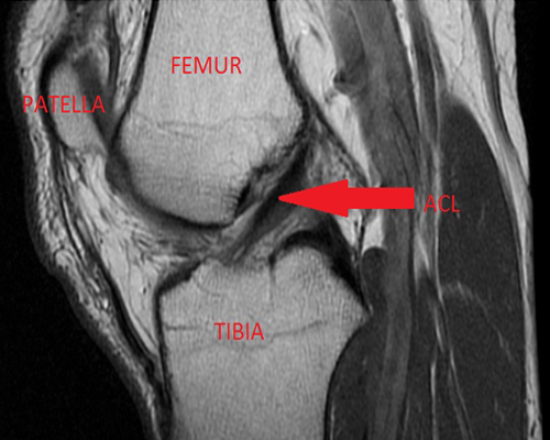
Figure 2: MRI image of the knee from the side and highlighting the anterior cruciate ligament.
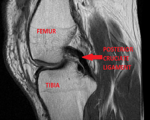
Figure 3: MRI image of the knee from the side and highlighting the posterior cruciate ligament.
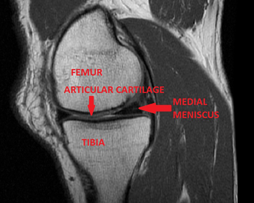
Figure 4: MRI image of the knee from the side and highlighting the medial meniscus.
Knee conditions
Below is a list and brief definition of some common knee conditions that will receive physiotherapy. There are many other knee conditions that are not listed. To receive a diagnosis, you should seek advice from a trained healthcare professional.
Osteoarthritis
This occurs through damage to the joint surface which means it does not move as well and is painful. It can be caused for a variety of reasons such as previous knee injury or surgery, from late 40's onwards, being overweight, being female or your parents had it. Symptoms that you can experience with osteoarthritis include pain on movement, stiffness after resting, swelling, restricted movement, difficulties or inability to do normal everyday activities or sport, grinding of the joint, giving way of the knee and changes in the shape of the leg (knee bowing out).
Other ligament injuries
Damage to the soft tissues that connect two bones together. There is often a specific injury that results in a ligament tear. The amount of damage to the ligament can be graded: 1 (a small number of fibres torn with localised pain and swelling), 2 (a large number of fibres torn with more severe pain, swelling and difficulty walking ), 3 (complete tear or rupture with significant pain, swelling, difficulty walking and a sensation of giving way). Knee ligaments that can get injured are:
- Posterior cruciate ligamentis commonly injured through a direct blow to the front of the tibia or landing and over bending the knee.
- Medial or lateral collateral ligamentis often injured during sideway movements that over extend the knee or due to repetitive trauma that put a strain on the ligament.
Meniscal tears
These are tears in the crescent shaped fibrocartilages that are located between the tibia and femur on the medial (inside) and lateral (outside) aspects of the knee. Tears can occur when the knee is bent or during twisting and turning activities. Sometimes tears occur due to degeneration within the meniscus and an individual cannot recall a specific injury. How a meniscus tear is treated will depend on the type of tear and symptoms an individual is experiencing.
Patella (knee cap) dislocations
This occurs when the patella (knee cap) which is at the front of the knee slides or comes out of place, normally to the outer side of the knee. Patella dislocations frequently occur during rapid changes of direction or due to direct trauma to the patella. When the patella comes out of joint then the medial patellofemoral ligament can be torn.
Patella subluxation
This occurs when the patella which is at the front of the knee partially slides or partially comes out of place, normally to the outer side of the knee. The knee can feel like it is going to give way.
Patellofemoral joint pain
This is pain that occurs around or behind the patella (knee cap). It can be caused for a variety of reasons such as; bony shape and structure, wear and tear, repetitive loading, blunt trauma, muscle deconditioning, altered biomechanics or secondary to another knee condition.
« Back to Menu ‹ Previous Page Next Page ›